 Dental imaging and x-rays provide enhanced views of dental health.
The results of dental imaging and x-rays allow dentists to accurately diagnose your dental needs and create effective treatment plans. These tools provide enhanced views of your teeth, bones and surrounding tissue.
3D Dental Imaging
3D Dental Imaging (also known as Cone Beam Computed Tomography, or CBCT) allows your dentist to see more anatomy, and with more clarity than traditional film-based dental x-rays. The focused x-ray beam reduces scatter radiation, resulting in better image quality and a lower dose of radiation. With an enhanced visualization of your teeth, bones, and surrounding hard and soft tissue, your doctor will understand more about your diagnosis and treatment plan.
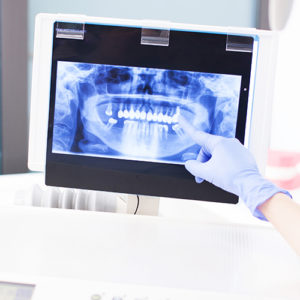
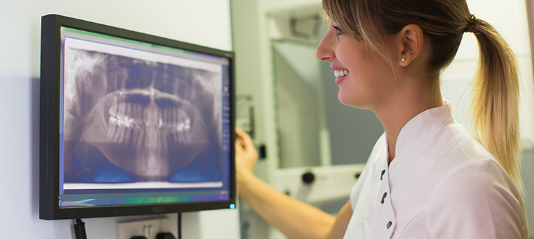
Panoramic X-rays
A panoramic x-ray is a commonly performed examination by dentists and oral surgeons in every day practice and is an important diagnostic tool. It covers a wider area than a conventional intraoral x-ray, and as a result, provides valuable information about the maxillary sinuses, tooth positioning and other bone abnormalities. This examination is also used to plan treatment for full and partial dentures, braces, extractions and implants.
Digital X-rays
Digital radiography (x-rays) benefits both the patient and the environment. One major benefit of digital radiography is that it uses 90% less radiation than conventional x-rays. The “film” used is actually a sensor that sends the x-ray image directly to a computer where the image is instantaneously displayed on a screen. Patients can view images on a large computer screen instead of the small, dark conventional x-ray film, which allows patients easier identification and education regarding any dental concerns. For a better diagnosis, these digital images can be magnified 300 times, made lighter or darker, can highlight specific areas and the contrast within the images can be clarified for sharpness. These images can be emailed to specialists for instant consultations or printed for patients to take home. There are no harmful processing chemicals to handle or dispose of, and there is no film to mount. Digital images will be stored on the computer, making paper charts obsolete.
Schedule your appointment today!